Case Study Series
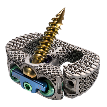
NanoHive Medical HiveTM Standalone ALIF System
Bridger Cox, MD
Neuroscience Specialists
Oklahoma City, OK
Technology Background
The design goals for NanoHive Medical’s Hive™ implant, with Soft Titanium® technology, are to offer a powerful feature set and several advatages over traditional PEEK or solid titanium interbody devices, including:
- A supporting scaffold for bony ongrowth
- Ideal pore characteristics for bony ingrowth
- A modulus of elasticity that is matched closely to cancellous bone
- Improved radiographic imaging
Bony Ongrowth:
As compared to PEEK, titanium surface textured implants have been demonstrated to enhance bone formations by increasing the osteogenic response and recruitment of mesenchymal stem cells (1).
A subtractive surface treatment is applied throughout the Hive implants to support osteoblast adhesion and optimize the environment for bony on-growth (2).
Bony Ingrowth:
Approximately 70% porous by volume, with 300-900 μm interconnected pores that have shown to be optimal for new bone attachment. Open pore structure allows for flow of cells and signals and the endplate-to-endplate channels are designed to promote bony ingrowth.
Cortical and trabecular bone growth was observed within the Soft Titanium lattice at 4 weeks. Bone growth was observed within the lattice before being observed in the packed lumen. Bone growth within the lumen is the only ingrowth observed in traditional PEEK implants (3).
Reduced Stiffness:
Hive interbodies using Soft Titanium technology have an elastic modulus similar to PEEK.
Patented rhombic dodecahedron lattice structure, similar to the base structure for cancellous bone, stimulates and and strengthens bone formation as described by Wolff’s Law.
Improved Radiographic Imaging:
Low density titanium structure improves radiolucency and reduces scatter on CT and MRI scans.
Study Summary
Sixty (60) patients (male – 33/60, female – 27/60) underwent Anterior Lumbar Interbody Fusions (ALIF) from October 2021 to November 2023 with the Hive Standalone ALIF Interbody System. The average patient age was 59.3 year old and the average BMI was 31.0. Nicotine use was recorded as Yes for 18/60 and No for 42/60.
A total of 103 levels were fused, including 58 at L5-S1, 29 at L4-L5, 10 at L3-L4 and 6 at L2-L3. An allograft bone graft was used by filling the central lumen and pores throughout the cage. Pedicle fixation was typically used as well, for additional support and stabilization.
Post-operative static x-rays were taken at 6 weeks, and 3 and 6 months, and assessed for fusion by observing for lucencies around the cage and screws. Additionally, CT scans were available for selective patients at 6 months.
Results
All patients were assessed at 6 months and determined to be either fused, when a CT scan was available, or radiographically progressing towards fusion with a stable construct and no lucencies around the interbody device or screws. There were no reoperations at the operative levels.
Conclusions
Dr. Cox has previously used allograft and standalone PEEK and titanium coated PEEK cages. Titanium cages have been an improvement in fusion rates. Hive Soft Titanium lattice, with the ability for bone graft to fill in and around the cage, has been a significant advantage. Available implant sizes fit the vast majority of patients, and the 15 degree lordosis option is particularly beneficial to restore and maintain sagittal balance. Instruments are intuitive and straightforward, making it efficient to implant and fixate with screws and locking plates. No revisions for pseudarthrosis have been reported and early solid bridging bone at 6 months is consistently observed.
Patient #1
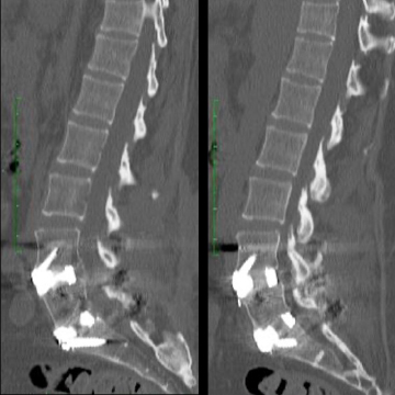
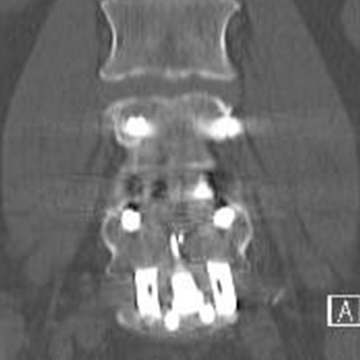
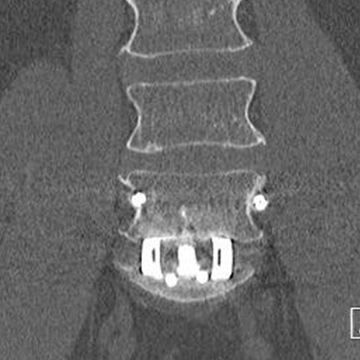
58 year old male, with a two level, L4-S1, ALIF
—
Six (6) month post-operative CT scan
—
Demonstrated fusion with solid bridging bone through the interspace
6 Months
6 Months
6 Months
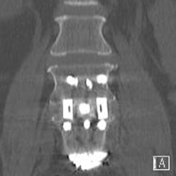
6 Months
Patient #2
A 54 year old male, with a two-level, L4-S1, ALIF
—
Six (6) month post-operative CT scan
—
Demonstrated fusion with solid bridging bone through the interspace
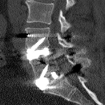
6 Months
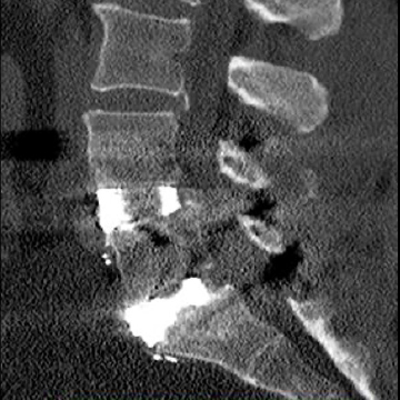
6 Months
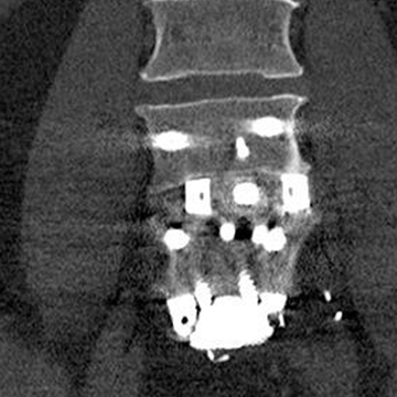
6 Months
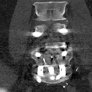
6 Months
Patient #3
A 56 year old female, L5-S1, ALIF
—
Six (6) month post-operative CT scan
—
Demonstrated fusion with solid bridging bone through the interspace
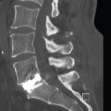
6 Months
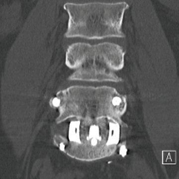
6 Months
Patient #4
A 63 year old female, L5-S1, ALIF
—
Six (6) month post-operative CT scan
—
Demonstrated fusion with solid bridging bone through the interspace
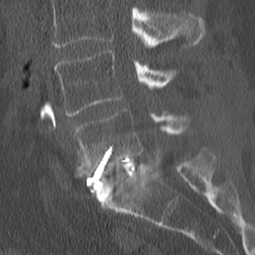
6 Months
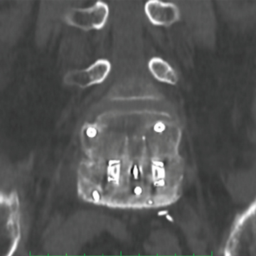
6 Months
2. Bassous, N, J., et al., 3-D printed Ti-6Al-4V scaffolds for supporting osteoblast and restricting bacterial functions without using drugs: Predictive equations and experiments, Acta Biomaterialia, 2019. vol 96: p. 662-673.
3. Jones, Bichara, et al. “Bone In Growth with 3D-Printed Soft Titanium® Scaffold.” [White Paper], NanoHive Medical, LLC. 2018.
