FOUR PILLARS
OF FUSION SUCCESS
HiveTM bioactive titanium interbody devices deliver all the benefits of a 3D printed titanium spine implants with none of the drawbacks typically associated with some of the more traditional designs.
The Nanohive Difference
Learn about the NanoHive difference from our Co-Founder and VP of Research and Development, Ian Helmar, and hear how the rhombic dodecahedron lattice provides biomechanical advantages for the Hive spine implants.
SOFT TITANIUM® TECHNOLOGY
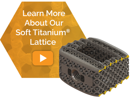
Unlike most titanium spine implants on the market, Hive interbody fusion devices were designed from the inside out, by first starting with NanoHive’s proprietary Soft Titanium® lattice as the foundation for the Hive portfolio. Because of its unique, energy absorbing rhombic dodecahedron design, the biomimetic lattice is durable enough to support biomechanical loading without the need for solid, stress shielding elements.
Durable Load Bearing Lattice
- Reduced stiffness of titanium lattice structure – effective modulus comparable to PEEK
- Rhombic dodecahedron lattice similar to the base structure for cancellous bone
- Maintains all structural strength requirements for cyclic biomechanical loading scenarios [1]
- Lack of solid stress shielding elements with independent endplates and a loadbearing core reduces overall implant stiffness
Comparison of Effective Modulus

DRIVING BONE ONGROWTH
NanoHive’s highly specific, sub-micron sized features on the spine implants’ surface are shown to interact with cells on a molecular level, kickstarting a biological cascade that supports adhesion of osteoblasts and drive bone attachment and formation.
- Complex sub-micron surface increases osteoblast proliferation [6,8]
- Increased osteoblast recruitment compared to untreated surfaces [6]
- Rapid bone attachment at endplates and throughout lattice [7]
- Soft Titanium® lattice provides up to 20x more surface area [1]
- Faster implant stability from on-growth at host bone interface [6]

ENHANCED BONY INGROWTH
The NanoHive Soft Titanium® Lattice provides the optimal balance of strength and porosity for bony ingrowth and its open pore structure allows for flow of cells and signals and interdigitation of bone growth throughout the Hive spine implants.
Ideal Pore Characteristics Promote Rapid Bone Infiltration [7]
- Pore sizes of 300 to 900 microns shown to be optimal for new bone attachment
- 70% porous, allowing bony ingrowth
- Lattice observed to promote bone growth prior to a lumen packed with autograft
- 22.1% volumetric bone formation at 4 weeks as compared to 8.9% in PEEK
Bone In Growth

A rabbit study shows ingrowth into Soft Titanium® and PEEK samples at 4 weeks and 8 weeks. Substantial growth into Soft Titanium® devices as compared to PEEK [7]
Lapine Model: 8 Weeks Data on File
NanoHive titanium lattice implants, packed with autograft, were implanted into a rabbit distal femoral condyle. At 8 weeks, the implant was explanted, and micro-CT cross sections, as seen in the below image, were observed. The 8-week micro-CT image demonstrates substantial bone growth within and adjacent to the implant. [7]

SUPERIOR IMAGING
The low density NanoHive Soft Titanium® Lattice allows for superior imaging and assessment of fusion on x-ray images or CT scans. Surgeons do not need to compromise on imaging when using the Hive titanium cages.
Visualization Available Across All Devices
- Hive spine implants preferred by four out of five surgeons over competitive devices in an independent blind study of visualization characteristics. [9]
- Endplate fusion readily identifiable on x-ray
- Easily visualize bone growth through cage
- Minimal scatter on CT and MRI scans

NanoHive Medical, LLC (formerly HD Lifesciences, LLC) Virtual Patent Marking
The NanoHive Medical, LLC products listed below are protected by patents in the United States and elsewhere. This website is provided to satisfy the virtual patent marking provisions of various jurisdictions including the virtual patent marking provisions of the America Invents Act (September 16, 2011).
The following list of products may not be all inclusive, and any other product not listed here may be protected by one or more patents. The products listed may be protected by additional patents, and other patents may be pending in the U.S. and elsewhere.
– Hive C, TL, PL, AL & AL Standalone Interbody Systems
– Protected by U.S. Patents:
• 9,962,269; 10,405,983 (Implant with Independent Endplates) • 10,888,429; 10,368,997 (Three Dimensional Lattice Structure for Implants) • 10,695,184; 15/895,213 (Methods of Designing Lattice Structures) • 15/895,213 (Methods of Designing Lucent Structures) • 10,624,746 (Fluid Interface System for Implants) • 10,881,518 (Anisotropic Biocompatible Lattice Structure) • 16/523,962 (Cervical Stand Alone)
[1] Test data on file. [2] BONE HOLD SPACE [3] Christopher L. Jones, Bartosz Wojewnik, Jason Tinley. (2018). Achieving Total-Implant Bone-Matched Modulus with 3D-Printed Soft Titanium® in Spinal Interbody Fusion [White Paper]. HD Lifesciences. [4] https://www.matweb.com/search/datasheet_print.aspx?matguid=2164cacabcde4391a596640d553b2ebe [5] Materials Properties Handbook: Titanium Alloys, R. Boyer, G. Welsch, and E. W. Collings, eds. ASM International, Materials Park, OH, 1994. [6] N. J. Bassous, C. L. Jones and T. J. Webster, 3-D printed Ti-6Al-4V scaffolds for supporting osteoblast and restricting bacterial functions without using drugs: Predictive equations and experiments, Acta Biomaterialia 96 (2019) 662–673, https://doi.org/10.1016/j.actbio.2019.06.055 [7] CL Jones, D Bichara, J Toy, J Tinley. (2018) “Bone In Growth with 3D-Printed Soft Titanium® Scaffold.” [White Paper], HD LifeSciences, LLC. [8] Ejiofor J., et al, Bone Cell Adhesion on Titanium Implants with Nanoscale Surface Features. International Journal of Powder Metallurgy, 40(2), 43-53. HD Lifesciences. [9] Christopher L. Jones, Ilsa Webeck, Jason Tinley. (2018). NanoHive™ with Soft Titanium® is Preferred 3D Printed Interbody Device on Radiographic Inspection [White Paper]. HD Lifesciences.


